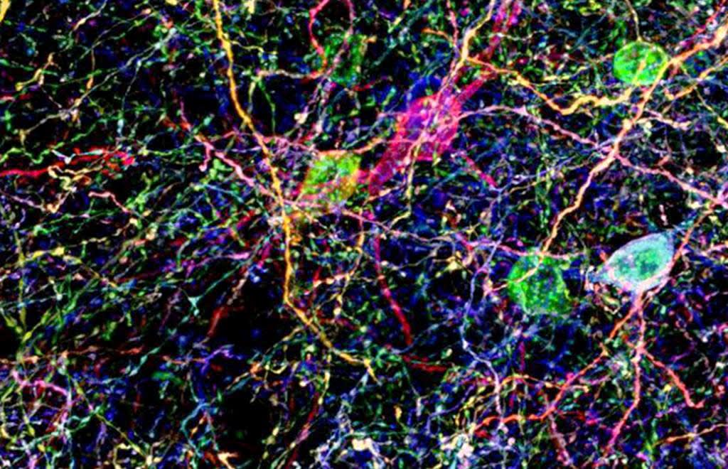
The research tool: Brainbow antibodies
Brainbow antibodies were generated by Dawen Cai, PhD, at University of Michigan, to primarily use as part of multicolour labelling strategy for fluorescent imaging of neuronal circuits and individual neurons in mice, Drosophila, and zebrafish and non-neuronal cells in mice.
The contributor
University of Michigan
Multicolour labelling strategy for fluorescent imaging
The Brainbow technology uses stochastic and combinatorial expression of fluorescent proteins to generate colour patterns, which serves as unique identification tag in cells and anatomically complex tissues, such as central nervous system. Dawen Cai, et al. (1) generated a refinement of the Brainbow technology (Brainbow 3) overcoming some of the limitations of the initial approach, such as suboptimal fluorescence intensity, failure to fill all axonal and dendritic processes, and disproportionate expression of the non-recombined fluorescent protein in the transgene. Brainbow 3 includes transgenic murine lines and adeno-associated virus (AAV). This version can also label delicate axonal and dendritic processes by using farnesylated derivatives of fluorescent proteins.

Brainbow applications*
- Lineage labelling of neurons and non-neuronal cells in mice, e.g., Confetti1 (2) and Ubow (3) models
- Cell tracing and lineage analysis in zebrafish, e.g., Zebrabow (4) and Drosophila, e.g., dBrainbow (5)
- Short-term cell labelling in different species via somatic expression, e.g., Brainbow AAV (5)
- Barcoding of somatic mutations and visualisation of clonal expansion and spread of oncogenes in the Crainbow mouse model (6)
Conclusion: Brainbow antibodies
- Custom-made polyclonal antibodies with specificity for eight different fluorescent proteins
- Fluorescent proteins derived from organisms of different species (except for EGFP and YFP) to increase specificity and avoid cross-reactivity
- Antibodies raised against different host species (e.g., chicken, rabbit, rat, and Guinea pig) to allow broader choice of secondary antibodies
- Antibodies can be used regardless of the sample species, as they react to the relevant fluorescent protein
- Use is recommended when the endogenous fluorescence of Brainbow is too weak, such as after fixation in histological analysis, to amplify the fluorescent signal
Explore the brainbow antibodies by the fluorescent proteins they target:
References
(1) Cai et al. 2013. Nat Methods. 10(6): 540–547. PMID: 23817127
(2) Snippert et al. 2010. Cell. 143(1):134-44. PMID: 20887898
(3) Ghigo et al. 2013. J Exp Med. 210(9): 1657–1664. PMID: 23940255
(4) Pan et al. 2013. Development. 140(13): 2835–2846. PMID: 23757414
(5) Hampel et al. 2011. Nat methods. 8(3): 253–259. PMID: 21297621
(6) Boone PG, et al. 2019. Nat Commun. 10(1):5490. PMID: 31792216
*List is not exhaustive