Category

Case-study

GATA3-eGFP reporter cell line: Advancing cancer drug discovery and Innovative tools – Insights from Matthew Holley

The research tool: GATA3-eGFP reporter cell line

GATA3, zinc finger transcription factor, is associated with numerous types of cancer in which its level of expression is critical. Drugs that modulate GATA3 expression are of particular interest, which is why a high-throughput  screening tool is an important addition to the research tool portfolio. Matthew Holley, Emeritus Professor at the University of Sheffield, has shared insights about such one-of-a-kind tool – the GATA3-eGFP reporter cell line, and other in vitro models  he has developed throughout his research- which he has generously contributed to CancerTools.org.

There are no other cell lines with this potential for the study of GATA3 in cancer research.

Emeritus Prof. Holley

The contributor

Emeritus Professor Matthew Holley

University of Sheffield; School of Biosciences

GATA3 in development and cancer

GATA3 is one of the GATA family transcriptional factors; zinc finger proteins that bind the consensus DNA sequence (T/A)GATA(A/G) of a target gene promoter. It is highly conserved among vertebrates and expressed in various tissues and cell lineages, such as immune cells, adrenal glands, placenta, kidneys, skin and breast tissue, inner ear, hair follicles, and nervous system.

GATA3 is responsible for regulating transcription during both development and cell differentiation, and its expression level is crucial at embryonic and postnatal stages. The importance is apparent from examples like early embryonic lethality due to GATA3 deletion, HDR syndrome associated with GATA3 haploinsufficiency, and the fact that Gata3 has been implicated in tumorigenesis. Previous studies have shown that it is involved in T-cell neoplasms, antagonizes cancer progression in PTEN-deficient prostates, and represents a useful marker for luminal category tumours in breast cancer.

GATA3-eGFP reporter cell line for drug discovery

In 2009, Matthew’s group published  results from a gene array analysis they conducted using inner ear cell lines [1].  They showed  a clear, previously unknown, functional link between GATA3 and the Akt signalling pathway; increased activity of which is often associated with tumour progression and therapies resistance. The pathway is essential for regulating cell survival and proliferation, and aberrations in its activation are linked to several human cancers, including breast, lung, ovarian, and prostate cancers.

Matthew delved into exploring how to modulate  gata3  expression, recognising its fundamental role in significant pathways associated with cancer progression as well as in the development of various organs and the nervous system.

Image: Inventing institution: University of Sheffield, UK

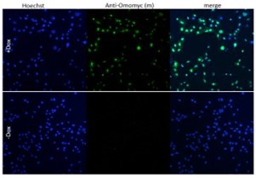
The GATA3-eGFP cell line was established by the cross of two well-characterised transgenic mouse lines. One mouse line stably expressed the H-2Kb-tsA58 transgene in which a temperature-sensitive variant of the SV40 large T-antigen was expressed under the control of an inducible promoter driven by gamma-interferon. The second, GATA3eGFP BAC-transgenic mouse line expressed eGFP under the control of the GATA3 promoter. The clones were selected according to the characteristics of the original tissue.

The GATA3-eGFP reporter cell line expresses GATA3-eGFP via an enhancer that is expressed in a pattern very closely resembling that of native GATA3 during mouse embryonic development. Fusion of GATA3 enhancer to eGFP is a key feature of this cell line allowing high-throughput screening of drugs that modulate the expression level of gata3.

First high content screening tests with established GATA3 modulators have shown effectiveness at the anticipated concentrations in the GATA3-eGFP cell line. The tool stands out as the only GATA3 reporter line capable of efficiently screening extrinsic factors influencing GATA3 expression in high-throughput systems.

The key objective of this case study is to show the high potential of this tool for those research laboratories interested in understanding GATA3’s role in various processes in cancer and other diseases.

Other in vitro models

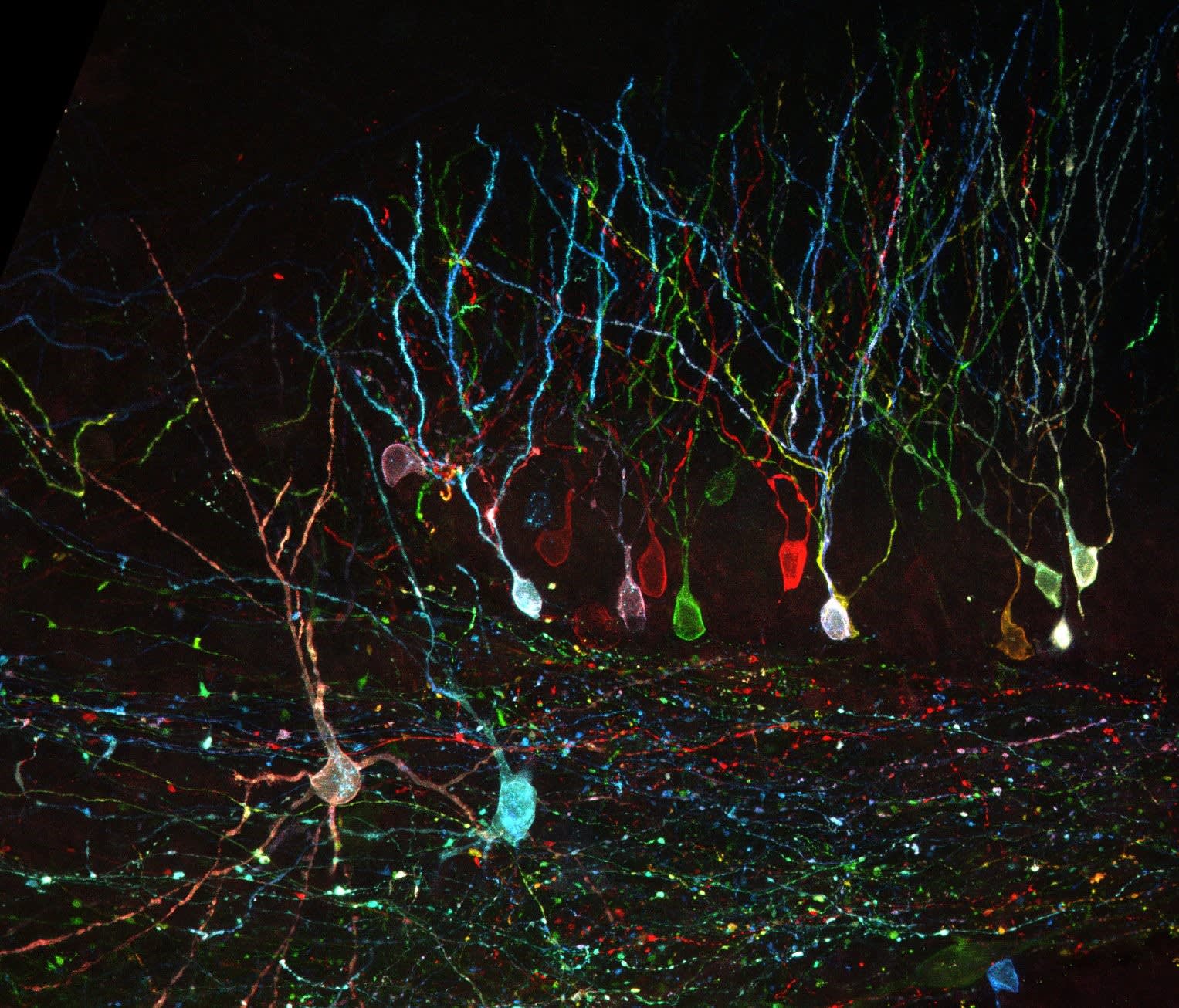
For many years, Matthew dedicated his research efforts to understand development and functioning of the inner ear in mammals, with an emphasis on exploring potential therapies for hearing loss.

Mammalian hair cells located in the inner ear are sensory cells that detect sound, gravity, and acceleration. Progressive loss of the hair cells is one of the main causes of deafness. Hearing loss is widespread and mostly irreversible as the mammalian cochlea is unable to regenerate hair cells that are lost, unlike amphibians and birds.

Mammalian auditory research is complicated by the fact that the very small numbers of auditory sensory epithelial cells do not proliferate postnatally or  in vitro,  which makes them experimentally inaccessible. Thus, Matthew and his colleagues developed a number of  in vitro  models for the differentiation of sensory hair cells and sensory nerves.

The established inner ear cell lines are derived from known cell populations during specific times of mouse inner ear development. They have been shown to correlate with gene expression profiles of the location from which they were derived. The lines are also conditionally immortal and can be grown under proliferating or differentiating conditions.

These cell lines were generously contributed to CancerTools.org and used for exploring the possibility of inner ear regeneration through both cell transplantation and the activation of early developmental processes [2, 3].

I believe in open access to all reagents where possible. They are costly to make and their application can advance research as well as return some investment back to the charitable funding agencies that enabled their production.

Emeritus Prof. Holley

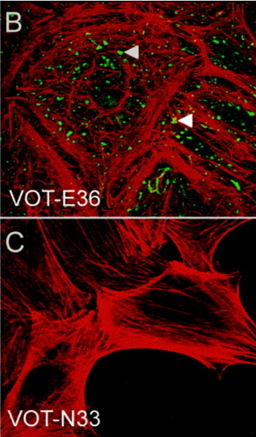
Image: Expression of the β4 integrin subunit in VOT-N33 and VOT-E36. (Adopted from Lawoko-Kerali et. al. 2004. Dev Dyn. 231(4):801-14. PMID: 15499550 Fig.2 B, C) 

Conclusion:

At CancerTools.org  we continue to work with  Cancer scientists who can deposit research tools they have developed in their labs, including cell lines, antibodies,  organoids, mouse models, cell culture media, small molecules and other state-of-the-art technologies, into our biorepository etc.

Access Emeritus Prof. Holley's cell lines:

References

1. Milo et.al. 2009. PLoS One. 4(9):e7144. PMID: 19774072.

2. Rivolta & Holley. 2002. J Neurobiol. 53(2):306-318. PMID: 12382283. 

3. Lawoko-Kerali et. al. 2004. Dev Dyn. 231(4):801-814. PMID: 15499550. 

Targeting the difficult-to-drug protein – MYC

The research tool: Anti-Omomyc

Cancer is associated with the abnormal growth of cells which proliferate uncontrollably and can metastasize into surrounding tissues. There are over 200 types of cancer and distinct subtypes have been identified. Adding to the challenging complexities of cancer, is tumour heterogeneity, which refers to the differences among tumours of the same type within different patients, among cells within a single tumour, or between a primary and secondary tumour (1). This variability often reduces the efficacy of existing therapies, compromising patient outcomes.

The biotechnological breakthroughs of recent years pushed clinical trials and cancer treatment paradigm to progressively shift from tumour-type centred to precision cancer medicine (2), where the focus is on patient-specific therapies, and challenges like tumour heterogeneity can be overcome through the development of specialized treatments. In contrast, discoveries at the Vall d’Hebron Institute of Oncology (VHIO) support the possibility of a more universal approach, with a single therapeutic option potentially targeting different types of cancer. By taking a universal approach to cancer therapy and targeting the common drivers of cancers across cancer types, this could result in therapies that are universal to cancer patients, but also specific to cancerous cells. The difficulty with this approach, is that these universal targets are often harder to drug, either due to the location or interactions of the target, or because of the universal target’s role in normal cellular function.

CancerTools.org spoke to Dr. Laura Soucek, an investigator at the VHIO and ICREA-born spin-off Peptomyc S.L., and leader of VHIO’s Models of Cancer Therapies Group to find out more.

The contributor

Dr. Laura Soucek

VHIO, ICREA-born spin-off Peptomyc S.L., and leader of VHIO’s Models of Cancer Therapies Group

Targeting a protein altered in many cancer types

Dr. Soucek entered research with one goal in mind – to make a difference in cancer research. To that end, she decided to focus on targeting a protein that was, and still is, considered by many to be “difficult-to-drug” – MYC. The MYC protein sits within the nuclei and acts as a ‘master regulator’ of a variety of cellular functions, including proliferation, differentiation, metabolism, and survival. Due to its multidimensional role, it is critical for MYC to be tightly regulated. Deregulation of MYC leads to altered cellular proliferation and growth, protein synthesis, and metabolism. Additionally, MYC promotes tumour progression through activation of angiogenesis and suppression of the host immune system. However, MYC is a difficult target for cancer therapy. Its location within the nuclei, together with its intrinsically disordered structure and lack of a specific active site, make direct MYC inhibition with traditional strategies challenging. Moreover, all three MYC family members (MYC, MYCN, and MYC) need to be targeted to obtain the most efficient therapeutic impact.

A long road to success

For 20 years, Dr. Soucek has been researching ways to inhibit MYC. In 1998, she designed the dominant-negative form of MYC called Omomyc, as a laboratory tool to study MYC biology. Since then, many milestones have been achieved; first using in vitro systems, transgene expression of Omomyc inhibited MYC activity and reduced proliferation in normal and cancer cells (3). Later, in various animal models of cancer, MYC inhibition by Omomyc exerted remarkable anti-cancer properties, without adverse and irreversible effects (4). Following Omomyc’s successful characterisation as a MYC inhibitor, it is now undergoing clinical development.

When we turned off MYC in cancer cells using Omomyc, we saw that it had a dramatic therapeutic effect in different types of experimental cancer models. The beauty of it is that, while everybody expected normal proliferating cells to suffer too, they simply slowed down their proliferation, but nothing major happened to them. So, we finally had a tool against cancer that seemed not to cause any severe side effects in normal proliferating tissues. And the other thing that really made me happy was that it appeared to be the opposite of personalised medicine: We had a technology that would be applicable to all types of cancers, so that maybe we didn't need different drugs for each of them, maybe we could use a common one for all cancers and patients.

Dr. Soucek
Image: A549 cells with Doxycycline-inducible Omomyc were orthotopically implanted into recipient mice. After treatment with Doxycycline, lung tissue was collected and FFPE lung sections were stained with anti-Omomyc monoclonal [21-1-3].

Omomyc impact

After the promising results obtained in vitro and in vivo, the next step was the conversion of Omomyc to an administrable drug. Dr. Soucek and her team showed that, as an alternative to its use as a transgene, Omomyc could be produced as a recombinant mini-protein. This purified Omomyc mini-protein then showed in Dr.Soucek’s studies that it spontaneously penetrates cancer cells and effectively interferes with MYC transcriptional activity, both in vitro and in vivo (5).

The first Omomyc-derived compound, OMO-103, successfully completed a phase 1 clinical trial in 2022. Previous results, presented at the 34th EORTC-NCI-AACR Symposium, Barcelona, 26-28 October 2022, showed that this first-in-class MYC inhibitor had few side effects, was tolerable, and stabilized disease in some patients. Patients in the trials had a range of solid tumours including pancreatic, bowel, and non-small cell lung cancers, and had received at least three prior lines of therapy (6).

Lasting structural integrity of therapeutic proteins in the target tissue is crucial to maintain their proper function. Since the in vivo stability of these agents can be affected by proteolytic degradation, the validity of Omomyc as the first direct inhibitor of MYC has frequently been questioned (8). A recently published study demonstrated, for the first time, that Omomyc behaves as a stable protein in tumour tissue, with longer lasting structural integrity compared to in blood (7).

Our findings are especially relevant now that Omomyc is being evaluated in clinical trials (Ph1/2 trial) since pharmacokinetic data are usually collected by analysis of blood samples in clinical practice,

Dr. Soucek

Conclusion:

A study published in the journal Genes & Development showed that the expression of Omomyc in preclinical melanoma models disrupts MYC activity and alters gene expression profiles, reducing cancer proliferation and progression (10). This suggests that MYC-targeted therapy by Omomyc could potentially open up a new treatment avenue for melanoma and point to the future development of clinical trials to assess the efficacy of this mini-protein against this tumour type (8).

In addition to the Omomyc mini-protein, Dr. Soucek’s laboratory also developed a monoclonal antibody targeting Omomyc, which can be used to study Omomyc levels, as well as better understand its function and effect on biological processes of interest, including cancer.

Omomyc: promising results seen in clinical trials

Discover Dr. Soucek's anti-Omomyc:

Brainbow Antibodies

The research tool: Brainbow antibodies

Brainbow antibodies were generated by Dawen Cai, PhD, at University of Michigan, to primarily use as part of multicolour labelling strategy for fluorescent imaging of neuronal circuits and individual neurons in mice, Drosophila, and zebrafish and non-neuronal cells in mice.

The contributor

Dr. Dawen Cai

University of Michigan

Multicolour labelling strategy for fluorescent imaging

The Brainbow technology uses stochastic and combinatorial expression of fluorescent proteins to generate colour patterns, which serves as unique identification tag in cells and anatomically complex tissues, such as central nervous system. Dawen Cai, et al. (1) generated a refinement of the Brainbow technology (Brainbow 3) overcoming some of the limitations of the initial approach, such as suboptimal fluorescence intensity, failure to fill all axonal and dendritic processes, and disproportionate expression of the non-recombined fluorescent protein in the transgene. Brainbow 3 includes transgenic murine lines and adeno-associated virus (AAV). This version can also label delicate axonal and dendritic processes by using farnesylated derivatives of fluorescent proteins.

Brainbow applications*

  • Lineage labelling of neurons and non-neuronal cells in mice, e.g., Confetti1 (2) and Ubow (3) models
  • Cell tracing and lineage analysis in zebrafish, e.g., Zebrabow (4) and Drosophila, e.g., dBrainbow (5)
  • Short-term cell labelling in different species via somatic expression, e.g., Brainbow AAV (5)
  • Barcoding of somatic mutations and visualisation of clonal expansion and spread of oncogenes in the Crainbow mouse model (6)
Image: Neurons and interneurons labelled with Brainbow AAV injected into cortex of PV-Cre mice. Antibody amplified mTFP and EYFP are in green, TagBFP is in blue and mCherry is in red. From: Cai et al. 2013. Nat Methods. 10(6):540-7. PMID: 23817127.

Conclusion: Brainbow antibodies

  • Custom-made polyclonal antibodies with specificity for eight different fluorescent proteins
  • Fluorescent proteins derived from organisms of different species (except for EGFP and YFP) to increase specificity and avoid cross-reactivity
  • Antibodies raised against different host species (e.g., chicken, rabbit, rat, and Guinea pig) to allow broader choice of secondary antibodies
  • Antibodies can be used regardless of the sample species, as they react to the relevant fluorescent protein
  • Use is recommended when the endogenous fluorescence of Brainbow is too weak, such as after fixation in histological analysis, to amplify the fluorescent signal

Explore the brainbow antibodies by the fluorescent proteins they target:

References

(1) Cai et al. 2013. Nat Methods. 10(6): 540–547. PMID: 23817127
(2) Snippert et al. 2010. Cell. 143(1):134-44. PMID: 20887898
(3) Ghigo et al. 2013. J Exp Med. 210(9): 1657–1664. PMID: 23940255
(4) Pan et al. 2013. Development. 140(13): 2835–2846. PMID: 23757414
(5) Hampel et al. 2011. Nat methods. 8(3): 253–259. PMID: 21297621
(6) Boone PG, et al. 2019. Nat Commun. 10(1):5490. PMID: 31792216

*List is not exhaustive

The importance of studying Non-Melanoma Skin Cancer

The research tool: Diagnostic research tools

Professor Leigh and her team have contributed to various streams of SCC and BCC research, focusing mainly on keratinocyte biology; ranging from epithelial differentiation and the cell biology of SCC, to the molecular machinery driving non-melanoma skin cancer. Professor Leigh was a pioneer in these fields, entering research at a time when keratinocyte growth was a new technology, and was involved with producing various important antibodies and cell lines in BCC and SCC cancer identification.

We spoke with Professor Leigh and her team to delve into the importance of this stream of cancer research and what the contribution of tools can do for SCC and BCC.

The contributor

Professor Irene Leigh

Queen Mary University of London; CRUK, London Research Institute: Lincolns Inn Fields; CRUK, London Research Institute: Clare Hall Laboratories

Non- Melanoma Skin Cancer (NMSC's) diagnostic research tools

Non-melanoma skin cancers (NMSCs or Keratinocyte cancers-KC) including basal cell carcinoma (BCC) and squamous cell carcinoma (SCC),  receive less awareness than their melanoma counterpart. This is because they are perceived as causing fewer deaths, as only SCCs metastasise. However, the incidence of NMSC is very high (18-20 times higher than that of melanoma) and there are 156,000 new non-melanoma skin cancer cases in the UK every year (2016-2018) – almost 430 every single day. Non-melanoma skin cancers commonly develop due to the skins exposure to the sun. Because these cancers are caused by excessive ultraviolet radiation, patients often end up having more than one skin cancer, resulting in multiple surgeries. Both types of carcinoma have an excellent prognosis, are slow-growing and rarely metastasise, however, in some cases they can develop into invasive skin cancers, with an aggressive nature.

It is critical to study NMSCs to produce diagnostic tests which identify high-risk tumours for metastasis and progression and generate treatments which limit carcinoma recurrence, as this is more challenging to treat.

Basal Cell Carcinoma (BCC) and Squamous cell Carcinoma (SCC)

One group of patients that are significantly affected by SCC and BCC are the immunosuppressed. With over 40% of SCC mortalities occurring in organ transplant receivers, this group has an important impact on immunotherapy treatment options and survivability. In working with these patients, Professor Leigh and her team have established a unique panel of patient-derived cutaneous squamous cell carcinoma cell lines. One example from this panel is the MET1 SCC cell line.

The development of MET1 SCC cell line

This cell line was derived from a primary lesion on the hand of an immunosuppressed patient, which ultimately recurred (MET2 cell line) and metastasised (MET4SCC line), representing crucial stages in SCC transformation. Understanding the stages a squamous cell carcinoma takes in order to become increasingly invasive helps dictate preventative and therapeutic measures for this cancer. This cell line has also been used in a variety of research; from indicating HPV isn’t essential for cancerous phenotype maintenance, to furthering our understanding of epithelial-mesenchymal transition within metastasis.

Recessive Dystrophic Epidermolysis Bullosa (RBDEB)

RDEB is a debilitating condition involving a deficiency in anchoring fibrils,  predominantly type VII collagen – between the epidermis basement membrane and underlying connective tissue. This leads to extremely fragile skin with severe blistering, meaning most patients develop SCC before the age of 35. Irene and her team helped to uncover the genetic basis of this condition through their research.

The development of Anti-Collagen Type VII LH7.2 antibody

Anti-Collagen Type VII LH7.2 is a monoclonal antibody which binds to an epitope of type VII collagen within the basement membrane of stratified squamous epithelia. This antibody helps diagnose RDEB as LH7.2 binding is absent or significantly reduced in RDEB patients. LH7.2 can also be used for differentiating invasive from non-invasive melanoma by assessing the integrity of epidermal basement membranes. This enables us to predict how aggressive SCC might be in RDEB patients.

The development of Anti-Katine 5/6/18 [LP34]

Professor Leigh and her team also created a monoclonal antibody that could be used to diagnose SCC and BCC tumours. Keratins 5, 6 are members of the type II keratins family that are specifically expressed in the inner root sheath of hair follicles. Keratins demonstrate tissue and differentiation-specific expression profiles. The type II cytokeratins consist of basic or neutral proteins which are arranged in pairs of heterotypic keratin chains co-expressed during differentiation of simple and stratified epithelial tissues. Keratin 18 is a member of the type I intermediate filament chain keratin 18. Keratin 18, together with its filament partner keratin 8, are perhaps the most commonly found members of the intermediate filament gene family. These cytokeratins have been reported to be expressed in tumour cells of epithelial origin and less commonly of mesothelial origin- however, non-epithelial tumours, e.g. lymphomas, do not express these cytokeratins. Anti-Katine 5/6/18 LP34 identifies tumours with an epithelial origin and is key in pathological diagnosis and understanding metastases. As this antibody binds to keratins 5, 6 and 18, it can also be used to identify uncultured keratinocyte material. Polyspecific LP34 also has an uncommonly broad pattern of reactivity, staining all human epithelial cells – both stratified and simple epithelium.

The impact

Irene Leigh and her lab have made significant contributions to the understanding and treatment of non-melanoma skin cancers and skin diseases, generating a number of cancer research tools that can be used to diagnose these conditions. Despite the belief that these cancers are not a danger due to their high treatability and low mortality levels, investigating them can have far-reaching impacts. Understanding the common pathways between NMSCs and keratinocytes can give insight into other cancers and skin conditions. Alongside this, studying these skin cancers can lead to new novel diagnostics and therapeutics to improve cancer survival rates within at-risk populations. By contributing these research tools to the initiative, researchers globally can now utilise these tools to continue to accelerate discoveries and help grow our understanding around non-melanoma skin cancers.

Not only do we need to be able to diagnose patients more accurately, we also need to search for clues for their treatment. This could come from a variety of sources, but you need the right reagents to do so

Professor Irene Leigh

Access the research tools:

[/vc_row_inner]

About CancerTools.org

CancerTools.org, the research tools arm of Cancer Research UK, is a non-profit, global community of cancer researchers, academic institutes and societies, with a shared mission to accelerate cancer research discoveries. In this collaborative, researchers contribute research tools and share knowledge to deepen our understanding of cancer, and drive innovation within cancer research.

About Prof. Irene Leigh

Professor Irene Leigh is currently a professor of Cellular Molecular Medicine at Barts and the London School of Medicine and Dentistry, Queen Mary University London. Leigh established the Centre for Cutaneous Research at Barts and The London School of Medicine and Dentistry, Queen Mary University of London (BLSMD) which went on to become the leading research centre in skin biology and disease in the United Kingdom.

The Kinase Chemogenomic Set (KCGS)

The research tool: The Kinase Chemogenomic Set (KCGS)

There are approximately 518 kinases encoded within the genome but for over 150 of these, their substrate is unknown, leaving much of the kinome poorly annotated and little understood with respect to its role in human biology.

Dysregulation of kinases is a leading cause of oncogenesis and disease including immune, neurological, and infectious ailments. Disruption of kinase activity, be it upregulation or deactivation, can impact cell survival, proliferation or migration. However, 90% of the current research effort has been expended on only 20% of known kinases.

Understanding the remaining unstudied ‘dark’ kinases is essential to provide opportunities to discover new drug targets and diagnostic tools. Yet, there is still a lack of research tools/resources dedicated to these kinases.

The contributor

Tim Willson

University of North Carolina at Chapel Hill (UNC) & Structural Genomics Consortium (SGC)

Developing a new tool kit

The Structural Genomics Consortium (SGC) is a non-profit organisation and home to scientists from medicinal chemistry, cell biology, and chemical biology. The SGC recognised the need to focus on the chemical biology of protein kinases. Operating out of the UNC Eshelman School of Pharmacy, these researchers have combined their skills to develop small molecule inhibitors aimed at ‘dark’ kinases to  understand their structure, substrate, and function. This work resulted in the creation of the Kinase Chemogenomic Set (KCGS).

Introducing the Kinase Chemogenomic Set (KCGS)

The Kinase Chemogenomic Set (KCGS) is the most diverse and highly-annotated, publicly-available collection of kinase inhibitors. Version 1.0 of the set contains 187 kinase inhibitors with potent activity on 215 human kinases, sourced from eight pharma companies and leading academic laboratories. Version 2.0 has currently replaced version 1.0, which features 108 additional kinase inhibitors, for a total of 295 kinase inhibitors.

KCGS helps scientists identify medically important kinases and allows for the synthesis of high-quality chemical probes for high-priority dark kinases of therapeutic interest. Each inhibitor has been cross-screened across hundreds of kinases – only those meeting strict selectivity criteria are included in the set.

The use of these chemical inhibitors alongside screening assays will help uncover the biology of these unknown/dark proteins in both health and disease. Preliminary characterisation of the KCGS in phenotypic screens showed its potential for chemogenomic exploration of kinase signaling, which could lead to new targets for drug discovery and precursors to new medicines.

KCGS impact

The KCGS set can be used in oncology and other life science research. A number of research projects have already made use of the KCGS to reduce the manual labour of sourcing and cross-screening each inhibitor across hundreds of kinases. Here are some of their results:

greatly benefited from using KCGS in investigating cellular mechanisms of drug resistance in cancer by the application of chemogenomic libraries to discover novel kinase targets for pancreatic cancer treatment. KCGS has opened up new programs to study the viral-induced kinome and the discovery of small molecule inhibitors as potential anti-viral drugs

Lee Graves, UNC School of Medicine

Patrick Eyer, Institute of Integrative Biology

Using the KCGS has shaken up investigation into the phosphoproteome. Patrick Eyers from the Institute of Integrative Biology was studying how covalent inhibitors of EGFR family protein kinases induce degradation of human Tribbles 2 (TRIB2) pseudokinae in cancer cells [3].

KCGS has changed how we think about targeting unusual protein kinases, including pseudokinases and conformationally-restricted canonical kinases, with small molecules

Patrick Eyer, Institute of Integrative Biology

Daniel Ebner, University of Oxford

Using the KCGS, Daniel Ebner, has been able to identify kinase targets in neurodegeneration, cancer, wound healing, inflammation and cardiovascular disease [1], leading to several manuscripts already in publication. For example, thanks to KCGS, a novel synthetic lethal interaction between cyclin F loss and Chk1 inhibition was discovered, paving the way for patient selection in the clinical use of checkpoint inhibitors [2]

Discover how you can benefit from KCGS:

References

[1] Wells, CI. et al. 2021. Int J Mol Sci. 22(2):566. PMID: 33429995
[2] Burdova, K. et al. 2019. EMBO J. 38(20):e101443. PMID: 3142411
[3] Foulkes, D.M. et al. 2018. Sci Signal. 11 (549): eaat7951. PMID: 30254057.

PlasmaxTM vs DMEM the impact of physiologically relevant cell culture media

The research tool: Plasmax

Choosing an appropriate cell culture medium is a crucial step in in vitro cell biology research. With a wide variety of media currently available, finding the correct one for your cell type and particular experiment can be challenging.

Sunada Khadka, a PhD Candidate at MD Anderson Cancer Center, studies cancer metabolism in glioma cells. Glioma is an intra-axial brain tumour which originates in the glial cells that surround and support neurons in the brains. During her latest research on anaplerosis in glioma cells, Sunada’s initial results obtained in vitro using traditional medium were not reproduced in her in vivo experiments. This led to additional time and resources being used to try and understand the discrepancy.

Here, we explore Sunada’s latest research, and the role PlasmaxTM, a physiologically relevant media, played in resolving the discrepancy between her in vitro and in vivo experimental results.

The researcher

Sunada Khadka

PhD candidate, MD Anderson Cancer Center

Initial in vitro results

Sunada’s research explored the possibility of synergistically killing tumour cells through the inhibition of glycolysis and glutaminolysis, two metabolic pathways that feed The Citric Acid (TCA) cycle.

A novel enolase inhibitor, HEX, was used as a glycolysis inhibitor in this study. HEX was developed through the concept of collateral lethality wherein the passenger deletion of the glycolytic gene ENO1 within a subset of gliomas, selectively renders cancer cells sensitive to inhibition of the redundant isoform ENO2. HEX was tested in combination with CB-839. CB-839 is a glutaminase inhibitor which targets glutamine metabolism and is currently being investigated in randomised clinical trials against a range of malignancies. This made CB-839 of primary interest to extend the metabolism-targeted therapy.

Initially, a pyruvate-free traditional media (DMEM) was used for the in vitro experiments which suggested a very strong effect of CB-839 on ENO1-deleted cancer cells. The combination of CB-839 and HEX provided a dramatic synergetic effect that seemed specific to ENO1-deleted cells.

Difficulty recapitulating in vitro results in an in vivo setting

However, when it was attempted to recapitulate the in vitro results in vivo, within an intracranial tumour model, no effect with CB-839 alone and no additive effects with HEX could be seen.

As CB-839 is known to be very poorly permeable across the brain, a subcutaneous in vivo tumour model was used, where Blood Brain Barrier penetration is not an issue. In this case some delay in tumour growth was observed after using CB-839 alone and when used in combination with HEX, but not to the extent seen in the in vitro research.

It was disappointing as we did not see any effect at all after the glutaminase inhibitor and that was very surprising because we saw a very dramatic effect - the complete wipe-out of cells - in vitro.

Sunada Khadka

Figure 1. ENO1-deleted glioma cells (D423) were implanted intracranially in immunocompromised nude mice and tumor growth was monitored weekly across different treatment groups by T2 MRI (indicated by dashed yellow outlines) 20-30 days after tumor implantation. Khadka et al. 2021.

Plasmax impact

This inconsistency in data led to a return to in vitro experimental conditions and a closer examination of the cell culture media used. PlasmaxTM was selected as a cell culture media that better reflected the in vivo nutrient profile. PlasmaxTM is a ready-to-use, physiologically relevant cell culture medium, consisting of >80 components, of which >50 have been optimised to levels found within human plasma.

By comparing the  in vitro results from PlasmaxTM to DMEM, it was observed that the toxicity of CB-839 in ENO1-deleted cells is significantly reduced in PlasmaxTM compared to DMEM. These results confirmed the in vivo data and demonstrated that the ENO1-deleted gliomas microenvironment may not be conducive to glutamine addiction.

We decided to try something that matches the physiological profile. And again we saw what we did not expect, which is that the effect of CB-839 seem to be completely diminished in PlasmaxTM medium compared to pyruvate-free DMEM.

Sunada Khadka

Figure 2. Sensitivity of glioma cells to CB-839 is attenuated in physiological Plasmax medium. ENO1 homozygously deleted (D423), ENO1-isogenic rescue (D423 ENO1), and ENO1 wild type (LN319) cells were grown in pyruvate free DMEM or Plasmax medium with or without 5 mM pyruvate supplementation. Khadka et al. 2021.

Conclusion:

Sunada’s results emphasize the importance of triaging your cell culture media with physiologically relevant media like PlasmaxTM to better recapitulate the in vivo environment.

As an extension of the paper she recently published, Sunada is now studying the effect of the glycolysis inhibitor in combination with an angiogenesis inhibitor. The restriction of oxygen and nutrient flow into the tumour should have a profound effect. For these experiments she plans on using an intracranial in vivo tumour model and together with PlasmaxTM in her in vitro experiments.

In the future, whatever metabolism related work I do, I'll make sure to compare DMEM to PlasmaxTM to ensure that the nutrient profile is not effecting the certain phenotype that I’m seeing. It doesn't hurt - if you are already doing one experiment in one certain media condition, just make another plate with PlasmaxTM for side-by-side comparison. So, I actually don't see why one wouldn’t try it. Especially before you jump into big in vivo experiments, which involve a lot of time and money. Using physiologically relevant media is a time saver and will make you more confident in your data.

Sunada Khadka

PlasmaxTM is already being repeatedly purchased by various cancer researchers across different academic institutes worldwide. 

Discover how PlasmaxTM could benefit your research:

About CancerTools.org

CancerTools.org, the research tools arm of Cancer Research UK, is a non-profit, global community of cancer researchers, academic institutes and societies, with a shared mission to accelerate cancer research discoveries. In this collaborative, researchers contribute research tools and share knowledge to deepen our understanding of cancer, and drive innovation within cancer research.

About Dr. Saverio Tardito

Dr. Saverio Tardito is the  group  leader for the oncometabolism research group at the CRUK Glasgow Beatson Institute and senior lecturer for the School of Cancer Sciences at the University of Glasgow.

About Cancer Research UK Glasgow: The Beatson Institute 

One of Cancer Research UK’s core-funded institutes, The Beatson Institute have built an excellent reputation for basic cancer research, including world-class metabolism studies and renowned in vivo modelling of tumour growth and metastasis. Learn more at:  https://www.beatson.gla.ac.uk/About/about-beatson.html

PlasmaxTM: A physiologically relevant cell culture media

The research tool: PlasmaxTM

PlasmaxTM is a physiologically relevant cell culture medium that closely resembles the metabolic and nutritional profile of human plasma. Unlike traditional media designed to supply excessive levels of a few nutrients, it provides unmatched metabolic fidelity.

CancerTools.org spoke with Dr. Tardito about PlasmaxTM to explore the importance of its development and what its contribution to the CancerTools.org initiative can do for cancer research.

The contributor

Dr. Saverio Tardito

Cancer Research UK Glasgow: The Beatson Institute

The future of cell culture media

Cell culture media is a critical component of cell-based assays, but its contribution to results is often overlooked. The right cell culture is critical in order to comprise the correct energy and compounds to regulate and support the cell cycle. For in vitro experiments, scientists typically use media like Dulbecco’s Modified Eagle Medium (DMEM) – a mixture of vitamins, selected amino acids, sugars and salts which sustain cellular growth. Yet, such types of mediums typically focus on cell proliferation rather than the nutritional environment that cells withstand in tumours. Dr. Tardito, an oncometabolism expert from the CRUK Beatson Institute, required a cell culture medium which better reflected human physiological conditions in order to study cancer biology.

Overcoming in vitro and in vivo variance

Traditional cell culture media was originally produced to rapidly and successfully increase cell proliferation in an in vitro environment (Eagle, 1955). This was made possible by adding nutrients in excessive concentrations to avoid nutrient depletion and simultaneously promote cell growth. Such disproportionate nutrient composition, in comparison to in vivo conditions like human plasma, affects both phenotypic and genotypic behaviour of cells (Schug et al, 2015 and Tardito et al, 2015). Usage of traditional media for cell culture can therefore lead to unrepresentative in vitro conditions and variance between in vitro and in vivo cancer cell metabolism. This becomes particularly important in research relating to cancer cell biology and related metabolic pathways.

To address this challenge, the research team at the Beatson Institute for Cancer Research, Glasgow, UK, under the supervision of Dr. Saverio Tardito, developed a novel cell culture medium, PlasmaxTM, to study the cell metabolism in different tumour types.

The clear cut differences between experiments performed with Plasmax vs commercial media available at the time become obvious once you realise, they aren’t physiologically relevant

Dr. Tardito

The development of PlasmaxTM

Dr. Tardito and his research team, optimised the concentrations of over 80 compounds typically found in human plasma to achieve the cell growing conditions. The cell culture medium contains all relevant elements to mimic human plasma, consisting of proteinogenic amino acids, vitamins, salts, and sugars, determined through Dr.Tardito’s optimisation experiments. The inclusion of metabolites enhances its physiological relevance and thereby mimics the in vivo environment. Trace elements, while essential for survival and proliferation, are often missing from traditional cell culture media and have to be supplemented before use. PlasmaxTM, is uniquely formulated with trace elements including vanadium, zinc, manganese, copper and selenium. The presence of these increase the antioxidant capacity of cells, which promotes colony growth by preventing ferroptosis-induced cell death (VandeVoorde et al., 2019).

Benefits of PlasmaxTM

It is critical for biomedical research to renew and refine models to improve their relevance to human physiology – which is exactly what the development of PlasmaxTM helps to execute.

PlasmaxTM has been successfully validated across primary cells of different tissue, species, and experimental conditions (see Table 1), and is suitable for both primary and established cell lines. Additional cell lines are successfully cultured using PlasmaxTM  regularly, which makes Table 1 a running list of validated cell lines. PlasmaxTM is anticipated to work across a broad range of cancer cell culture models.

Using a physiological relevant medium significantly impacts the results obtained from common cellular assays, including colony formation and gene expression. This has the potential to improve results for cancer cell biology experiments associated with drug discovery and in vitro cancer models.

Using physiologically relevant media is a time saver and will make you more confident in your data.

Sunada Khadka

Physiologically relevant

PlasmaxTM is optimised to reflect the in vivo profiles of nutrients and metabolites found in human plasma, including essential and non-essential amino acids, amino acid derivatives, organic acids, and other polar metabolites.

Improves in vitro metabolic fidelity

PlasmaxTM can better approximate the overall metabolic phenotype of tumours, with both 2D and 3D cells cultured in PlasmaxTM, better recapitulating the tumours’ metabolic signatures.

By increasing the metabolic fidelity and biological relevance of in vitro cancer models, better drug discovery and improved understanding of cancer at a cellular level can ensure.

Produces a faster proliferation

When compared with traditional mediums, PlasmaxTM produces a faster proliferation, even when aged up to 12 months, in comparison with DMEM when both are supplemented with 2.5% foetal bovine serum.

Better mimics tumour metabolism

PlasmaxTM better mimics tumour metabolism. Breast cancer spheroids grown in PlasmaxTM , have shown to better approximate the metabolic profile of mammary tumours (Vande Voorde et al., 2019).

Uncover role of trace elements

Cancer cells seeded at low densities in the absence of the trace element selenium are unable to form colonies in traditional media due to lipid peroxidation and ferroptosis. The growth-enabling trace elements in addition to vitamins and inorganic salts in PlasmaxTM, prevent ferroptosis-induced cell death, and promote colony growth.

Table 1: A selected list of cultured cell lines successfully validated for growth and viability in PlasmaxTM under standard conditions

Cell lines grown in Plasmax TM Tissue of origin Cell line status Species
HepG2 Liver Cancer Established line Human
HuH7 Liver Cancer Established line Human
HuH6 Liver Cancer Established line Human
BT549 Breast Cancer Established line Human
MDA-MB-468 Breast Cancer Established line Human
Cal120 Breast Cancer Established line Human
A375 Melanoma Established line Human
Colo829 Melanoma Established line Human
LN18 Brain Cancer Established line Human
Naive glioblastoma cell line Brain Cancer Low passage lines Human
Dermal fibroblasts Epidermis Primary Human
Small intestine organoid Small intestine Primary Mouse
Mammospheres Mammary gland Primary Mouse
Mesenchimal stromal cell line Bone marrow Primary Human
Embryonic stem cell line Embryo Primary Human
Trophoblast stem cell line Placenta Primary Human
A549 Lung cancer Established line Human
HCT116 Colon Cancer Established line Human
SaOS2 Bone tumour Established line Human
HT1080 Fibrosarcoma Established line Human

Plasmax impact

Choosing an appropriate cell culture medium is a crucial step in in vitro cell biology research and finding the correct one for your cell type and experiment can be challenging. Such was experienced by Sunada Khadka, a PhD Candidate at MD Anderson, during her research on anaplerosis in glioma cells.

While using a traditional medium, Sunada’s initial results obtained in vitro were not reproduced in her in vivo experiments. This inconsistency in data led to a return to  in vitro  experimental conditions and a closer examination of the cell culture media used. PlasmaxTM  was selected as a cell culture media that better reflected the  in vivo  nutrient profile. By comparing in vitro results from PlasmaxTM to DMEM, Sunada was able to understand the discrepancy, illuminating the importance of triaging cell culture media with physiologically relevant media like PlasmaxTM  in order to better recapitulate the  in vivo  environment.   

In the future, whatever metabolism related work I do, I'll make sure to compare DMEM to Plasmax to ensure that the nutrient profile is not effecting the certain phenotype that I’m seeing

Sunada Khadka

Conclusion:

Optimising the relevant components in culture media and defining their physiological concentrations at scale, can be a challenging process. Having PlasmaxTM in a pre-prepared liquid form saves time, effort, initial investment of sourcing, and spares the process of optimising 80+ components to get their proportions accurate.   

PlasmaxTM unique formula, maintains its effectiveness throughout its shelf life with no effect on cell growth from being aged. It is compatible across different cell types and is greatly beneficial to any cancer researcher interested in the study of cancer cell biology, in vitro cancer models and cell based assays.

PlasmaxTM is already being repeatedly purchased by various cancer researchers across different academic institutes worldwide. 

Meet your research needs simply and accessibly:

About CancerTools.org

CancerTools.org, the research tools arm of Cancer Research UK, is a non-profit, global community of cancer researchers, academic institutes and societies, with a shared mission to accelerate cancer research discoveries. In this collaborative, researchers contribute research tools and share knowledge to deepen our understanding of cancer, and drive innovation within cancer research.

About Dr. Saverio Tardito

Dr. Saverio Tardito is the  group  leader for the oncometabolism research group at the CRUK Glasgow Beatson Institute and senior lecturer for the School of Cancer Sciences at the University of Glasgow.

About Cancer Research UK Glasgow: The Beatson Institute 

One of Cancer Research UK’s core-funded institutes, The Beatson Institute have built an excellent reputation for basic cancer research, including world-class metabolism studies and renowned in vivo modelling of tumour growth and metastasis. Learn more at:  https://www.beatson.gla.ac.uk/About/about-beatson.html

Introducing the pcPPT-mPGK-attR-sLPmCherry-WPRE vector

The research tool: pcPPT-mPGK-attR-sLPmCherry-WPRE

One of the major challenges for tumour research is identifying how tumour cells exploit their surrounding cells, the so-called tumour microenvironment (TME), to help form and expand the tumour itself.

Existing methods can be used to investigate this challenge, but all have disadvantages; for example, immunohistochemistry allows you to view the location of the TME cells but only in fixed samples, whilst flow cytometry allows the detection of different cell types within a tissue, but doesn’t show spatial information to specifically identify TME cells (Frontiers in Oncology 2018, 00390). A major advance in this field, would be to be able to specifically isolate the cells that have been corrupted, to understand the exact nature of how these cells were hijacked to support tumour progression.

The contributor

Ilaria Malanchi and Luigi Ombrato

The Francis Crick Institute

Facing the challenge

Ilaria Malanchi and Luigi Ombrato, researchers at The Francis Crick Institute, were working on this challenge. They were growing frustrated with the lack of available tools to isolate cells surrounding the tumour mass at the point of tumour corruption. They decided a new system was needed to tackle this challenge of identifying cells within the TME, isolating them and then studying exactly what they are. Although both were aware of the challenges involved in this task and were dubious that they would even succeed, the need for the tool outweighed the challenges. Ilaria and Luigi took inspiration from different sources of literature and developed a system to engineer tumour cells, giving them the unique ability of labelling their surrounding cells. It took a lot of resilience, dedication and alterations, but after almost three years, they had a vector they thought could work; pcPPT-mPGK-attR-sLPmCherry-WPRE.

Introducing the pcPPT-mPGK-attR-sLPmCherry-WPRE vector

The pcPPT-mPGK-attR-sLPmCherry-WPRE vector is a breakthrough in tumour research. It allows for spatial identification of the local metastatic cellular environment within the whole tissue. Lentiviral transduction is used to introduce the vector to tumour cells which can then themselves be introduced to the study host. In vivo, these cells release the cell-penetrating fluorescent red protein mCherry, which is taken up by neighbouring cells within the TME. Once the tumour environment has developed, cells can be harvested and analysed by FACS. The results enable identification of cell types directly engaged by the tumour and comparison with remaining unlabelled cells from the same tissue. Malanchi and Ombrato have shared protocols on how to engineer this vector for different cell types, giving researchers a powerful tool to apply across a variety of research areas including cancer and immunology, among others. Application of this new tool has enabled the Malanchi lab to identify the unprecedented presence of cancer associated parenchymal cells (CAPs) within the lung metastatic environment of breast cancer (Nature, 2019; 572(7771): 603-608).

Accelerating cancer research through research tools

Before joining the Francis Crick Institute, Luigi had never considered depositing his research tools. However, through the Francis Crick Institute’s partnership with CancerTools.org, Ilaria and Luigi were able to quickly and efficiently deposit their vector.

Making our research tools available is what we are all aiming for in a way – Looking at the benefits that our findings and science can have. I think it’s really important that people who do research have the possibility to translate it, but they need support. Scientists don’t necessarily have the skills to translate their research by themselves - they need support to be able to do the next step.

Luigi Ombrato

Having been through the process of depositing their research tools, Luigi firmly believes in the importance of making their research tools available to other scientists.

The deposit process was a very smooth process. Our technology transfer team at the Institute were always available and were very proactive in coming to us to propose things. I didn’t know about CancerTools.org beforehand and wouldn’t have known to contact them without the technology transfer team

Luigi Ombrato

Conclusion: impact of the vector

Following the publication and translation of the pcPPT-mPGK-attR-sLPmCherry-WPRE vector in Nature, CancerTools.org has received  enquiries from researchers around the world for access to the research tool. In addition, 10 global working groups have been already using the vector in a variety of research areas including breast cancer metastasis and leukaemia, with results from some of these groups expected to be published shortly.

Discover more about the vector:

About CancerTools.org

CancerTools.org, the research tools arm of Cancer Research UK, is a non-profit, global community of cancer researchers, academic institutes and societies, with a shared mission to accelerate cancer research discoveries. In this collaborative, researchers contribute research tools and share knowledge to deepen our understanding of cancer, and drive innovation within cancer research.

Tool enquiry

Please ensure you use your organisation email address rather than personal where possible, as this helps us locate your organisation in our system faster.

CancerTools.org uses the contact information provided to respond to you about our research tools and service. For more information please review our privacy policy.